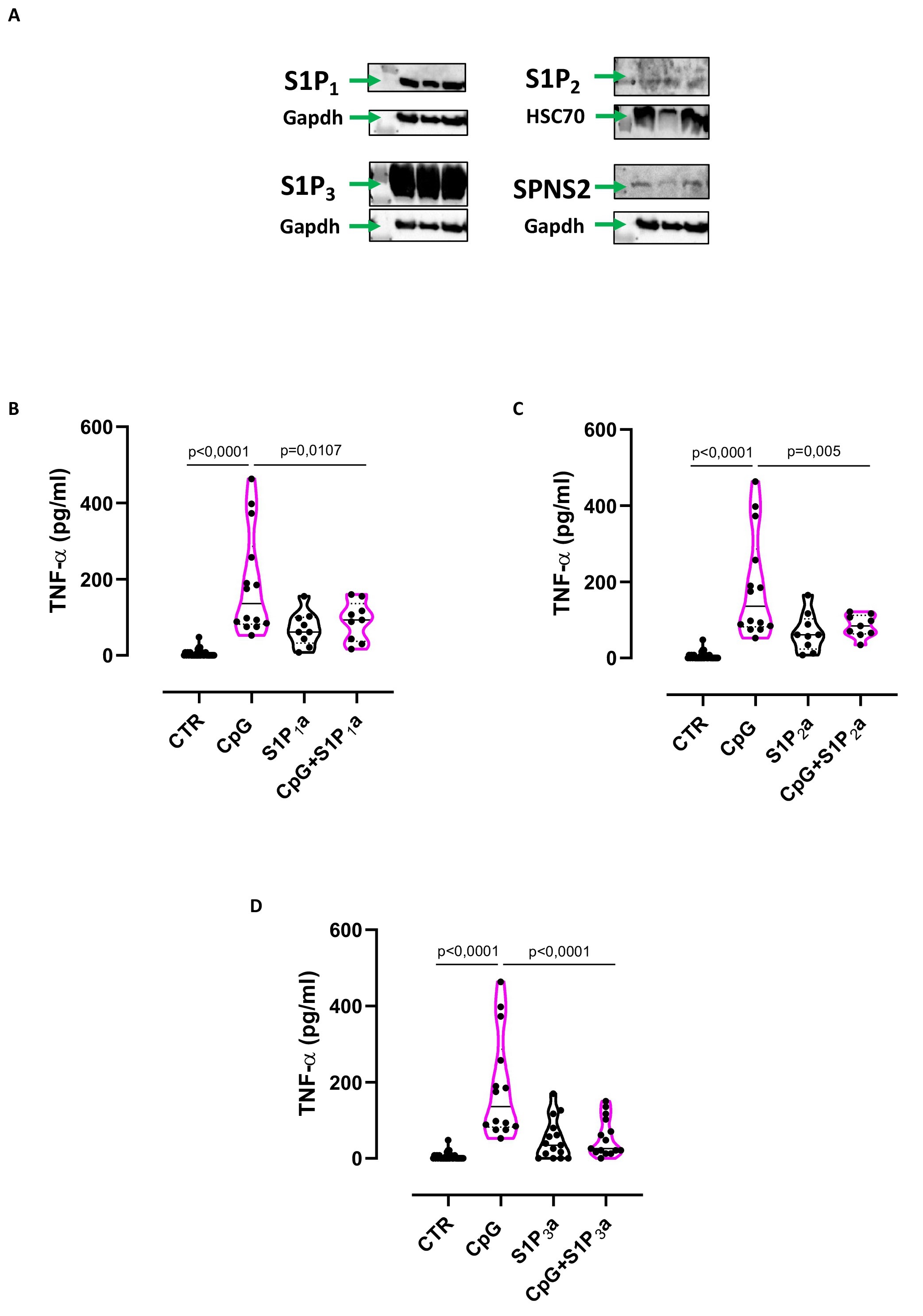
Fig. 3. TNF-α release mediated by TLR9/S1P-axis is S1P3 dependent. A) A549 expression of S1P1 (43kDa), S1P2 (42kDa), S1P3 (55-70kDa) and SPNS2 (70kDa) evaluated by western blotting assay. GAPDH and HSP70 were used as loading protein. Lung adenocarcinoma A549 cells were stimulated with TLR9 ligand, CpG 1 µg/ml for 8 hours and TNF-α release was evaluated after the inhibition of S1P receptors. The inhibition of both S1P1 (B), S1P2 (C) and S1P3 (D) significantly reduced the release of TNF-α after CpG stimulation. Data are presented as violin plot, showing the median ± interquartile range. Statistical differences were assessed with Ordinary ONE-Way ANOVA followed by Tukey's multiple comparison test. W146, S1P1 receptor antagonist (S1P1a, 1 µM), JTE-013, S1P2 receptor antagonist (S1P2a, 100 nM), TY52156, S1P3 receptor antagonist (S1P3a, 10 µM).